- Home
- Medical news & Guidelines
- Anesthesiology
- Cardiology and CTVS
- Critical Care
- Dentistry
- Dermatology
- Diabetes and Endocrinology
- ENT
- Gastroenterology
- Medicine
- Nephrology
- Neurology
- Obstretics-Gynaecology
- Oncology
- Ophthalmology
- Orthopaedics
- Pediatrics-Neonatology
- Psychiatry
- Pulmonology
- Radiology
- Surgery
- Urology
- Laboratory Medicine
- Diet
- Nursing
- Paramedical
- Physiotherapy
- Health news
- Fact Check
- Bone Health Fact Check
- Brain Health Fact Check
- Cancer Related Fact Check
- Child Care Fact Check
- Dental and oral health fact check
- Diabetes and metabolic health fact check
- Diet and Nutrition Fact Check
- Eye and ENT Care Fact Check
- Fitness fact check
- Gut health fact check
- Heart health fact check
- Kidney health fact check
- Medical education fact check
- Men's health fact check
- Respiratory fact check
- Skin and hair care fact check
- Vaccine and Immunization fact check
- Women's health fact check
- AYUSH
- State News
- Andaman and Nicobar Islands
- Andhra Pradesh
- Arunachal Pradesh
- Assam
- Bihar
- Chandigarh
- Chattisgarh
- Dadra and Nagar Haveli
- Daman and Diu
- Delhi
- Goa
- Gujarat
- Haryana
- Himachal Pradesh
- Jammu & Kashmir
- Jharkhand
- Karnataka
- Kerala
- Ladakh
- Lakshadweep
- Madhya Pradesh
- Maharashtra
- Manipur
- Meghalaya
- Mizoram
- Nagaland
- Odisha
- Puducherry
- Punjab
- Rajasthan
- Sikkim
- Tamil Nadu
- Telangana
- Tripura
- Uttar Pradesh
- Uttrakhand
- West Bengal
- Medical Education
- Industry
Panoramic X-rays Identify Osteoporosis Risk via Jaw Bone Density, Study Finds

Brazil: Evaluating bone mineral density (BMD) in the mandibular ramus using panoramic dental X-rays could serve as an indicator for identifying individuals at risk of osteoporosis, a recent study has found.The findings, published in the Journal of Oral and Maxillofacial Surgery, suggest that panoramic radiography may serve as an adjunctive tool for osteoporosis screening."These results...
Brazil: Evaluating bone mineral density (BMD) in the mandibular ramus using panoramic dental X-rays could serve as an indicator for identifying individuals at risk of osteoporosis, a recent study has found.
The findings, published in the Journal of Oral and Maxillofacial Surgery, suggest that panoramic radiography may serve as an adjunctive tool for osteoporosis screening.
"These results highlight the importance of panoramic radiography as a supplementary tool for detecting individuals susceptible to osteoporosis, particularly when evaluating BMD in regions such as the mandibular ramus, unaffected by dental artifacts and substantial muscular factors," the researchers wrote.
Osteoporosis, frequently seen in postmenopausal women, results in substantial bone density loss and increases fracture susceptibility. Cortical bone, the primary reservoir of calcium in the skeleton, is particularly vulnerable to conditions like osteoporosis. Panoramic radiography, which captures cortical bone in the lower jaw canal, can thus aid in evaluating BMD.
Against the above background, Angela Jordão Camargo, University of São Paulo, São Paulo, Brazil, and colleagues aimed to characterize and compare changes in the cortices of the mandibular canal between normal, osteopenic, and osteoporotic postmenopausal women.
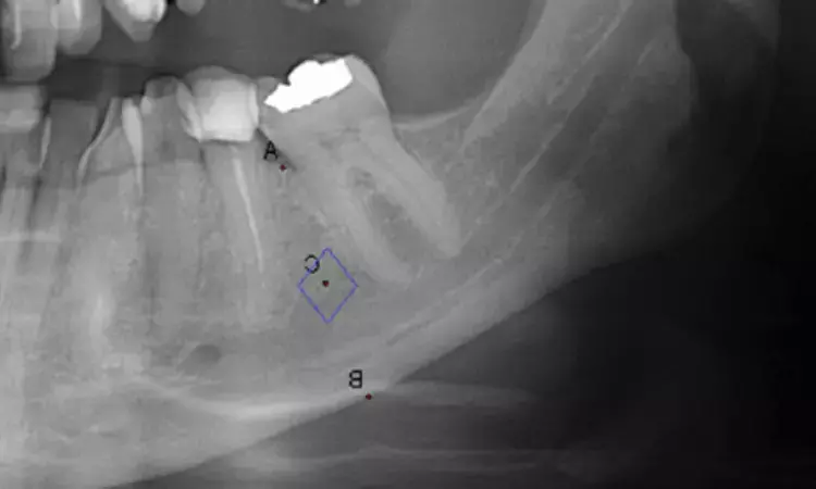
For this purpose, the researchers assessed the panoramic X-rays from 52 normal, osteopenic, and osteoporotic postmenopausal women over 40. All women underwent osteoporosis risk assessment by dual-energy x-ray absorptiometry (DEXA). In the cross-sectional study, the intensity of black pixels on radiographs was utilized to measure the BMD of the cortices of the mandibular canal.
The study led to the following findings:
· The sample comprised 52 postmenopausal women over 40 years, 26 normal, 18 osteopenic, and eight osteoporotic.
· Significant differences were observed in the percentage of black pixels in the mandibular ramus between the groups.
· In this region, the average percentage of black pixels was 3.19% for the normal group, 2.78% for the osteopenia group, and 2.35% for the osteoporosis group.
· There were no significant differences in other mandibular regions.
"Our findings show an association between BMD assessed in the mandibular canal cortex and the osteoporosis presence as determined by DXA," the researchers wrote.
"Although the differences in black pixel percentages observed in the mandibular ramus are subtle, they are statistically significant, indicating that panoramic radiography could be a valuable adjunctive tool for osteoporosis screening," they concluded.
Reference:
Camargo, A. J., Rodrigues, G. A., Munhoz, L., Lourenço, A. G., & Watanabe, P. C. A. (2024). Are radiographic changes in the mandibular canal associated with bone mineral density? Journal of Oral and Maxillofacial Surgery. https://doi.org/10.1016/j.joms.2024.06.167
Dr Kamal Kant Kohli-MBBS, DTCD- a chest specialist with more than 30 years of practice and a flair for writing clinical articles, Dr Kamal Kant Kohli joined Medical Dialogues as a Chief Editor of Medical News. Besides writing articles, as an editor, he proofreads and verifies all the medical content published on Medical Dialogues including those coming from journals, studies,medical conferences,guidelines etc. Email: drkohli@medicaldialogues.in. Contact no. 011-43720751


