- Home
- Medical news & Guidelines
- Anesthesiology
- Cardiology and CTVS
- Critical Care
- Dentistry
- Dermatology
- Diabetes and Endocrinology
- ENT
- Gastroenterology
- Medicine
- Nephrology
- Neurology
- Obstretics-Gynaecology
- Oncology
- Ophthalmology
- Orthopaedics
- Pediatrics-Neonatology
- Psychiatry
- Pulmonology
- Radiology
- Surgery
- Urology
- Laboratory Medicine
- Diet
- Nursing
- Paramedical
- Physiotherapy
- Health news
- Fact Check
- Bone Health Fact Check
- Brain Health Fact Check
- Cancer Related Fact Check
- Child Care Fact Check
- Dental and oral health fact check
- Diabetes and metabolic health fact check
- Diet and Nutrition Fact Check
- Eye and ENT Care Fact Check
- Fitness fact check
- Gut health fact check
- Heart health fact check
- Kidney health fact check
- Medical education fact check
- Men's health fact check
- Respiratory fact check
- Skin and hair care fact check
- Vaccine and Immunization fact check
- Women's health fact check
- AYUSH
- State News
- Andaman and Nicobar Islands
- Andhra Pradesh
- Arunachal Pradesh
- Assam
- Bihar
- Chandigarh
- Chattisgarh
- Dadra and Nagar Haveli
- Daman and Diu
- Delhi
- Goa
- Gujarat
- Haryana
- Himachal Pradesh
- Jammu & Kashmir
- Jharkhand
- Karnataka
- Kerala
- Ladakh
- Lakshadweep
- Madhya Pradesh
- Maharashtra
- Manipur
- Meghalaya
- Mizoram
- Nagaland
- Odisha
- Puducherry
- Punjab
- Rajasthan
- Sikkim
- Tamil Nadu
- Telangana
- Tripura
- Uttar Pradesh
- Uttrakhand
- West Bengal
- Medical Education
- Industry
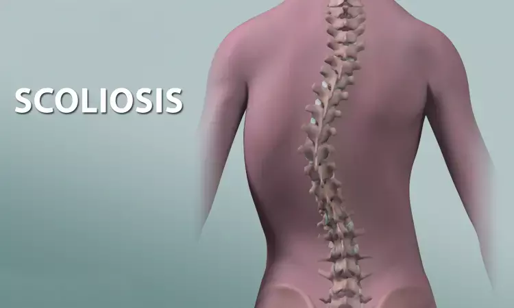
X-ray-like images from ultrasound scan through AI helpful for scoliosis diagnosis: Study

China: In a recent study, published in Ultrasonics, a team of researchers from China described the use of artificial intelligence (AI) algorithm that creates x-ray-like images from ultrasound data to visualize adolescent idiopathic scoliosis.
The results could be of significance for children at risk of scoliosis progression with their body growth, a condition known as adolescent idiopathic scoliosis (AIS) that affects 1% to 3% of the adolescent population. This technique could reduce radiation exposure and future cancer risk.
The gold standard for scoliosis diagnosis is standing X-ray radiograph with Cobb's method. However, its application gets restricted by radiation hazards, especially for close follow-up of adolescent patients. Ultrasound imaging compared with X-ray has the advantage of being radiation-free and real-time.
To combine the advantages of the above two imaging modalities described above, Cong Bai, College of Computer Science & Technology, Zhejiang University of Technology, Hangzhou, China, and colleagues proposed an ultrasound to X-ray synthesis generative attentional network (UXGAN) to synthesize ultrasound images into X-ray-like images. In this network, a cyclically consistent network was adopted and was trained end-to-end. The addition of an attention module was done and different residual blocks were designed.
The study included 202 children with adolescent idiopathic scoliosis -- all of whom underwent both ultrasound and x-ray. The quantitative comparison results showed the superiority of the method to the state-of-the-art CycleGAN methods. The researchers further compared the Cobb angle values measured on synthesized images and the real X-ray images, respectively. A good linear correlation (r = 0.95) was demonstrated between the two methods.
The researchers conclude, "the above results proved that the proposed method is of great significance for providing both X-ray images and ultrasound images based on the radiation-free ultrasound scanning."
According to the study authors, "the study findings show significant promise."
"[Our] experimental results show the reliability and accuracy of the method," they concluded. "This method can provide radiation-free ultrasound and x-ray images, which is of great significance for diagnosing and monitoring adolescent idiopathic scoliosis."
Reference:
The study titled, "Ultrasound to X-ray synthesis generative attentional network (UXGAN) for adolescent idiopathic scoliosis," was published in the journal Ultrasonics.
DOI: https://doi.org/10.1016/j.ultras.2022.106819
Dr Kamal Kant Kohli-MBBS, DTCD- a chest specialist with more than 30 years of practice and a flair for writing clinical articles, Dr Kamal Kant Kohli joined Medical Dialogues as a Chief Editor of Medical News. Besides writing articles, as an editor, he proofreads and verifies all the medical content published on Medical Dialogues including those coming from journals, studies,medical conferences,guidelines etc. Email: drkohli@medicaldialogues.in. Contact no. 011-43720751


