- Home
- Medical news & Guidelines
- Anesthesiology
- Cardiology and CTVS
- Critical Care
- Dentistry
- Dermatology
- Diabetes and Endocrinology
- ENT
- Gastroenterology
- Medicine
- Nephrology
- Neurology
- Obstretics-Gynaecology
- Oncology
- Ophthalmology
- Orthopaedics
- Pediatrics-Neonatology
- Psychiatry
- Pulmonology
- Radiology
- Surgery
- Urology
- Laboratory Medicine
- Diet
- Nursing
- Paramedical
- Physiotherapy
- Health news
- Fact Check
- Bone Health Fact Check
- Brain Health Fact Check
- Cancer Related Fact Check
- Child Care Fact Check
- Dental and oral health fact check
- Diabetes and metabolic health fact check
- Diet and Nutrition Fact Check
- Eye and ENT Care Fact Check
- Fitness fact check
- Gut health fact check
- Heart health fact check
- Kidney health fact check
- Medical education fact check
- Men's health fact check
- Respiratory fact check
- Skin and hair care fact check
- Vaccine and Immunization fact check
- Women's health fact check
- AYUSH
- State News
- Andaman and Nicobar Islands
- Andhra Pradesh
- Arunachal Pradesh
- Assam
- Bihar
- Chandigarh
- Chattisgarh
- Dadra and Nagar Haveli
- Daman and Diu
- Delhi
- Goa
- Gujarat
- Haryana
- Himachal Pradesh
- Jammu & Kashmir
- Jharkhand
- Karnataka
- Kerala
- Ladakh
- Lakshadweep
- Madhya Pradesh
- Maharashtra
- Manipur
- Meghalaya
- Mizoram
- Nagaland
- Odisha
- Puducherry
- Punjab
- Rajasthan
- Sikkim
- Tamil Nadu
- Telangana
- Tripura
- Uttar Pradesh
- Uttrakhand
- West Bengal
- Medical Education
- Industry
EBUS Offers Lifesaving Alternative for Loculated Pleural Collection After Lung Surgery: Case Report

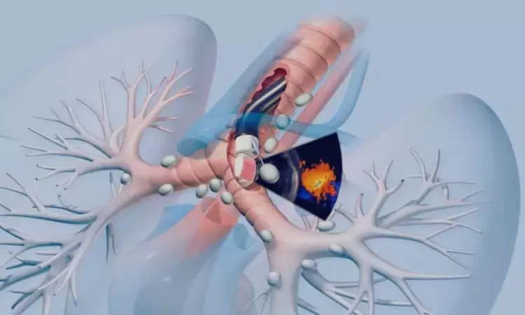
Turkey: A new case report published in BMC Surgery has demonstrated an innovative use of endobronchial ultrasound (EBUS) to diagnose a complex postoperative pleural collection in a patient with significantly altered thoracic anatomy.
The report, authored by Güntuğ Batıhan and İhsan Topaloğlu from the Department of Thoracic Surgery at Kafkas University, Turkey, describes how EBUS-guided aspiration provided a safe and minimally invasive alternative when conventional transthoracic approaches were deemed too risky.
Loculated pleural collections that develop after major lung surgery often resemble empyema and can be difficult to evaluate using standard methods. In this case, the surgical changes following right superior bilobectomy made access challenging, prompting clinicians to opt for a nontraditional approach. The patient, a 67-year-old man who had undergone surgery six months earlier for central squamous cell lung carcinoma, returned with clinical indicators of infection, including elevated inflammatory markers and leukocytosis.
Postoperative recovery for this patient had been complicated by a prolonged air leak and incomplete re-expansion of the remaining right lower lobe. Earlier imaging had shown a sterile, loculated collection in the upper right hemithorax. On his current presentation, CT scans revealed that this pocket had developed air–fluid levels suggestive of infection. However, due to mediastinal shift, obstruction by the scapula, and the close proximity of major vessels and ribs, conventional ultrasound-guided thoracentesis was considered unsafe.
Given the anatomical constraints, the team selected EBUS as a feasible alternative. After explaining potential risks such as bleeding and infection, they obtained informed consent and proceeded with the procedure under general anesthesia using a laryngeal mask airway. Through a right paratracheal route, the EBUS probe successfully identified the loculated pleural pocket. A 22-gauge EBUS-TBNA needle was then used to aspirate approximately 15 cc of pleural fluid, which appeared echogenic with visible debris.
While blood, urine, sputum cultures, and pneumococcal antigen testing showed no evidence of infection, microbiological analysis of the pleural fluid confirmed the presence of Streptococcus pneumoniae. The patient was treated initially with intravenous ceftriaxone for two weeks, followed by oral amoxicillin–clavulanate for an additional 14 days. He showed progressive clinical improvement, inflammatory markers normalized, and he was discharged in stable condition. At a nine-month follow-up, he remained symptom-free.
The authors emphasized that postoperative thoracic anatomy can significantly alter the feasibility of traditional diagnostic pathways. In this scenario, EBUS provided a practical and safe alternative for accessing pleural collections that otherwise could not be reached. They note that while EBUS is typically used for mediastinal lymph node evaluation, its application is expanding as clinicians recognize its potential in complex thoracic conditions.
The case highlights how interventional bronchoscopy techniques are evolving to meet diagnostic challenges in patients with surgically altered anatomy. As demonstrated here, EBUS-guided aspiration may offer a valuable option in selected cases where conventional approaches are limited or unsafe.
Reference:
Batıhan, G., Topaloğlu, İ. Transbronchial EBUS-guided aspiration of a loculated pleural collection following right superior bilobectomy: a case report. BMC Surg 25, 562 (2025). https://doi.org/10.1186/s12893-025-03317-6
Dr Kamal Kant Kohli-MBBS, DTCD- a chest specialist with more than 30 years of practice and a flair for writing clinical articles, Dr Kamal Kant Kohli joined Medical Dialogues as a Chief Editor of Medical News. Besides writing articles, as an editor, he proofreads and verifies all the medical content published on Medical Dialogues including those coming from journals, studies,medical conferences,guidelines etc. Email: drkohli@medicaldialogues.in. Contact no. 011-43720751
Next Story


