- Home
- Medical news & Guidelines
- Anesthesiology
- Cardiology and CTVS
- Critical Care
- Dentistry
- Dermatology
- Diabetes and Endocrinology
- ENT
- Gastroenterology
- Medicine
- Nephrology
- Neurology
- Obstretics-Gynaecology
- Oncology
- Ophthalmology
- Orthopaedics
- Pediatrics-Neonatology
- Psychiatry
- Pulmonology
- Radiology
- Surgery
- Urology
- Laboratory Medicine
- Diet
- Nursing
- Paramedical
- Physiotherapy
- Health news
- Fact Check
- Bone Health Fact Check
- Brain Health Fact Check
- Cancer Related Fact Check
- Child Care Fact Check
- Dental and oral health fact check
- Diabetes and metabolic health fact check
- Diet and Nutrition Fact Check
- Eye and ENT Care Fact Check
- Fitness fact check
- Gut health fact check
- Heart health fact check
- Kidney health fact check
- Medical education fact check
- Men's health fact check
- Respiratory fact check
- Skin and hair care fact check
- Vaccine and Immunization fact check
- Women's health fact check
- AYUSH
- State News
- Andaman and Nicobar Islands
- Andhra Pradesh
- Arunachal Pradesh
- Assam
- Bihar
- Chandigarh
- Chattisgarh
- Dadra and Nagar Haveli
- Daman and Diu
- Delhi
- Goa
- Gujarat
- Haryana
- Himachal Pradesh
- Jammu & Kashmir
- Jharkhand
- Karnataka
- Kerala
- Ladakh
- Lakshadweep
- Madhya Pradesh
- Maharashtra
- Manipur
- Meghalaya
- Mizoram
- Nagaland
- Odisha
- Puducherry
- Punjab
- Rajasthan
- Sikkim
- Tamil Nadu
- Telangana
- Tripura
- Uttar Pradesh
- Uttrakhand
- West Bengal
- Medical Education
- Industry
Noncontrast CT better alternative to CTA for predicting Hematoma expansion in ICH

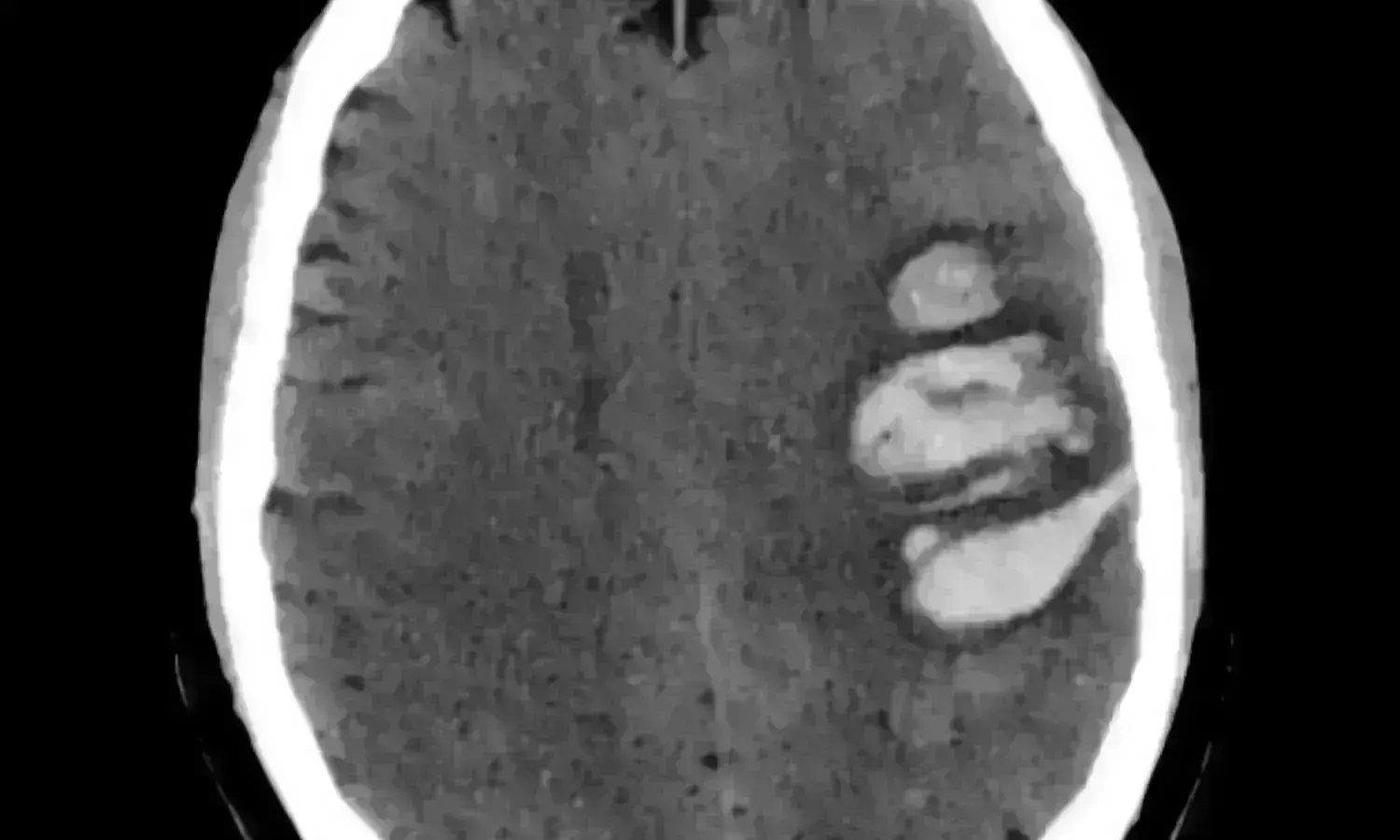
A new study found that in intracerebral hemorrhages for identifying hematoma expansion non contrast computed tomography hypodensities are better alternatives to CTA spot signs as non-contrast CT hypodensities significantly add value and predict hematoma expansion.
The study results are published in the journal Stroke.
Previous literature shows that in intracerebral hemorrhage non-contrast computed tomography hypodensities are a better predictor of hematoma expansion (HE) and a possible alternative to the computed tomography angiography (CTA) spot sign but there is uncertainty on the added value to available prediction models. Hence researchers conducted a multicenter study to investigate whether the inclusion of hypodensities improves the prediction of HE and compared their added value over the spot sign.
A multicenter, retrospective analysis was carried out at 8 university hospitals on patients admitted for primary spontaneous intracerebral hemorrhage.
| Boston | 1994-2015 | Prospective |
| Hamilton, Canada | 2010-2016 | Retrospective |
| Berlin, Germany | 2014-2019 | Retrospective |
| Chongqing, China | 2011-2015 | Retrospective |
| Pavia, Italy | 2017-2019 | Prospective |
| Ferrara, Italy | 2010-2019 | Retrospective |
| Brescia, Italy | 2020-2021 | Retrospective |
| Bologna, Italy | 2015-2019 | Retrospective |
Logistic regression analysis was used to explore the predictors of HE like the hematoma growth >6 mL and/or >33% from baseline to follow-up imaging. The discrimination of a simple prediction model for HE based on 4 predictors (antiplatelet and anticoagulant treatment, baseline intracerebral hemorrhage volume, and onset-to-imaging time) before and after the inclusion of non-contrast computed tomography hypodensities, were compared using the receiver operating characteristic curve and De Long test for the area under the curve comparison.
Key findings:
- A total of 2465 subjects were included, of whom 664 (26.9%) had HE and 1085 (44.0%) had hypodensities.
- After adjustment for confounders in logistic regression, hypodensities were independently associated with HE.
- The discrimination of the 4 predictors model improved with the inclusion of non-contrast computed tomography hypodensities.
- The added value of hypodensities was statistically significant but the addition of the CTA spot sign did not provide significant discrimination improvement in the subgroup of CTA patients (n=895, 36.3%).
Thus, non-contrast computed tomography hypodensities can be used to stratify the risk of HE with good discrimination without CTA.
Further reading: Morotti A, Boulouis G, Nawabi J, et al. Using Noncontrast Computed Tomography to Improve Prediction of Intracerebral Hemorrhage Expansion [published online ahead of print, 2023 Jan 9]. Stroke. 2023;10.1161/STROKEAHA.122.041302. doi: 10.1161/STROKEAHA.122.041302
BDS, MDS
Dr.Niharika Harsha B (BDS,MDS) completed her BDS from Govt Dental College, Hyderabad and MDS from Dr.NTR University of health sciences(Now Kaloji Rao University). She has 4 years of private dental practice and worked for 2 years as Consultant Oral Radiologist at a Dental Imaging Centre in Hyderabad. She worked as Research Assistant and scientific writer in the development of Oral Anti cancer screening device with her seniors. She has a deep intriguing wish in writing highly engaging, captivating and informative medical content for a wider audience. She can be contacted at editorial@medicaldialogues.in.
Dr Kamal Kant Kohli-MBBS, DTCD- a chest specialist with more than 30 years of practice and a flair for writing clinical articles, Dr Kamal Kant Kohli joined Medical Dialogues as a Chief Editor of Medical News. Besides writing articles, as an editor, he proofreads and verifies all the medical content published on Medical Dialogues including those coming from journals, studies,medical conferences,guidelines etc. Email: drkohli@medicaldialogues.in. Contact no. 011-43720751


