- Home
- Medical news & Guidelines
- Anesthesiology
- Cardiology and CTVS
- Critical Care
- Dentistry
- Dermatology
- Diabetes and Endocrinology
- ENT
- Gastroenterology
- Medicine
- Nephrology
- Neurology
- Obstretics-Gynaecology
- Oncology
- Ophthalmology
- Orthopaedics
- Pediatrics-Neonatology
- Psychiatry
- Pulmonology
- Radiology
- Surgery
- Urology
- Laboratory Medicine
- Diet
- Nursing
- Paramedical
- Physiotherapy
- Health news
- Fact Check
- Bone Health Fact Check
- Brain Health Fact Check
- Cancer Related Fact Check
- Child Care Fact Check
- Dental and oral health fact check
- Diabetes and metabolic health fact check
- Diet and Nutrition Fact Check
- Eye and ENT Care Fact Check
- Fitness fact check
- Gut health fact check
- Heart health fact check
- Kidney health fact check
- Medical education fact check
- Men's health fact check
- Respiratory fact check
- Skin and hair care fact check
- Vaccine and Immunization fact check
- Women's health fact check
- AYUSH
- State News
- Andaman and Nicobar Islands
- Andhra Pradesh
- Arunachal Pradesh
- Assam
- Bihar
- Chandigarh
- Chattisgarh
- Dadra and Nagar Haveli
- Daman and Diu
- Delhi
- Goa
- Gujarat
- Haryana
- Himachal Pradesh
- Jammu & Kashmir
- Jharkhand
- Karnataka
- Kerala
- Ladakh
- Lakshadweep
- Madhya Pradesh
- Maharashtra
- Manipur
- Meghalaya
- Mizoram
- Nagaland
- Odisha
- Puducherry
- Punjab
- Rajasthan
- Sikkim
- Tamil Nadu
- Telangana
- Tripura
- Uttar Pradesh
- Uttrakhand
- West Bengal
- Medical Education
- Industry
DMEK surgery significantly improves contrast sensitivity in Fuchs dystrophy: Study

Main surgical interventions in posterior corneal endothelial disorders, such as Fuchs endothelial dystrophy (FED) and bullous keratopathy (BK), are the posterior lamellar surgical techniques, Descemet stripping endothelial (automated) keratoplasty (DS(A)EK), and Descemet membrane endothelial keratoplasty (DMEK). The DMEK procedure results in better visual outcomes than the DS(A)EK procedure.
In the DMEK procedure, only an isolated Descemet membrane and its endothelium are transplanted. This can result in a near-normal anatomic corneal restoration. Visual outcome is mostly reported as best-corrected visual acuity (BCVA) under high-contrast conditions. The BCVA under high contrast conditions, however, is impaired in patients with posterior corneal endothelial disorders, and even other visual functions such as color vision and contrast sensitivity can be reduced.
Corneal aberrations are common in patients with posterior corneal endothelial disorders, and performing a DMEK procedure leads to changes of these corneal aberrations, which probably influences the contrast sensitivity.
In this study, authors Gundlach et al prospectively investigated preoperative and postoperative contrast sensitivities after DMEK and analyzed the influence of central corneal thickness (CCT) and anterior and posterior corneal aberrations on contrast sensitivities.
In this prospective study, 50 pseudophakic eyes of 50 patients who received DMEK surgery at the Charité—Universitätsmedizin Berlin were included. Visual acuity; contrast sensitivity using OPTEC 6500 at spatial frequencies of 1.5, 3, 6, 12, and 18 cycles/degree in photopic and mesopic light with and without glare; central corneal thickness (CCT); and anterior and posterior corneal aberrations were measured preoperatively and at 3 and 12 months postoperatively.
Best-corrected visual acuity (preoperative 0.67 ± 0.46 and after 12 months 0.19 ± 0.16 LogMAR, P < 0.001) and photopic and mesopic contrast sensitivities with and without glare improved significantly, whereas CCT decreased significantly (preoperative 677 ± 114 mm, after 12 months 527 ± 29 mm, P < 0.001).
Preoperative CCT correlates significantly with preoperative photopic contrast sensitivity (correlation coefficient 20.462, P = 0.002), and postoperative total anterior aberrations correlates with postoperative photopic contrast sensitivity (correlation coefficient 20.361, P = 0.006).
In this prospective observational case series, DMEK surgery improves the contrast sensitivities, the photopic and the mesopic with and without glare. Before surgery, authors found a significant correlation between contrast sensitivity and CCT, but no correlation is found between contrast sensitivity and anterior or posterior aberrations. After DMEK surgery, anterior corneal aberrations correlate to the contrast sensitivities, but there is no correlation between posterior corneal aberrations and contrast sensitivity.
Compared with healthy controls, contrast sensitivity in patients with FED or BK is significantly reduced. Especially, mesopic contrast sensitivity is inferior to photopic contrast sensitivity, and glare further reduces both mesopic and photopic contrast sensitivities. In this study, a significant improvement is evident after 3 months with an even further increase after 12 months.
Anterior and posterior corneal aberrations decrease after DMEK, but only anterior corneal aberrations (anterior total RMS), astigmatism of the anterior cornea, and values of the posterior keratometry show a significant decrease.
Analyzing the influence of CCT and anterior and posterior corneal aberrations on the contrast sensitivity, study correlated the contrast sensitivity to these factors. Authors found that there is a significant correlation between contrast sensitivity and CCT preoperatively, but no correlation is found between contrast sensitivity and anterior or posterior aberrations.
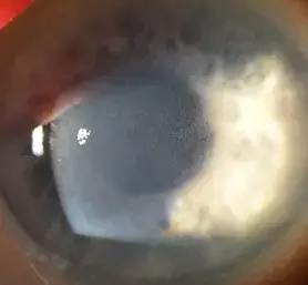
A thickened cornea and its associated corneal edema does not only lead to a massive scattering of the light but also affects the translucency of the cornea and consequently has an impact on the contrast sensitivity. Therefore, contrast sensitivity of patients with FED or BK seems to depend on the extent of the corneal thickness primarily. After DMEK, CCT normalizes, and there is no significant correlation between preoperative or postoperative CCT and postoperative contrast sensitivity anymore. Postoperative, anterior corneal aberrations demonstrate a high impact on contrast sensitivity, whereas posterior corneal aberrations do not correlate with contrast sensitivity.
In conclusion, photopic and mesopic contrast sensitivities, especially with glare, are preoperatively reduced in patients with FED and BK. The extent of the corneal thickening and the corneal edema seems to mainly influence the contrast sensitivity preoperatively. DMEK surgery significantly improves the contrast sensitivity. Postoperatively, contrast sensitivities are influenced by anterior corneal aberrations. Contrast sensitivity and its impact on patients' performance in everyday life situations should be considered when taking a decision whether a DMEK surgery should be performed.
Source: Gundlach et al; Cornea Volume 40, Number 9, September 2021
Dr Ishan Kataria has done his MBBS from Medical College Bijapur and MS in Ophthalmology from Dr Vasant Rao Pawar Medical College, Nasik. Post completing MD, he pursuid Anterior Segment Fellowship from Sankara Eye Hospital and worked as a competent phaco and anterior segment consultant surgeon in a trust hospital in Bathinda for 2 years.He is currently pursuing Fellowship in Vitreo-Retina at Dr Sohan Singh Eye hospital Amritsar and is actively involved in various research activities under the guidance of the faculty.
Dr Kamal Kant Kohli-MBBS, DTCD- a chest specialist with more than 30 years of practice and a flair for writing clinical articles, Dr Kamal Kant Kohli joined Medical Dialogues as a Chief Editor of Medical News. Besides writing articles, as an editor, he proofreads and verifies all the medical content published on Medical Dialogues including those coming from journals, studies,medical conferences,guidelines etc. Email: drkohli@medicaldialogues.in. Contact no. 011-43720751


