
SD-OCT Accuracy may help differentiate Papilledema from Pseudopapilledema, Suggests Study
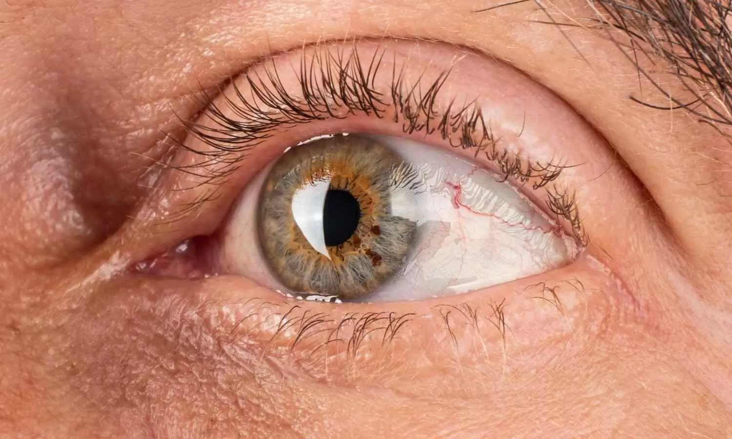


A recent cross-sectional case-control study published in the Journal of Clinical Ophthalmology and Research in April 2026 reveals that SD-OCT is a highly reliable, noninvasive tool for flawlessly distinguishing true optic disc swelling from its benign mimics. By measuring the average retinal nerve fiber layer thickness, it achieved near-perfect diagnostic accuracy with an impressive AUC of 0.997.
To safely distinguish life-threatening papilledema from benign pseudopapilledema without relying on unnecessary invasive procedures, Dr. Narayanan Balakrishnan's team analyzed SD-OCT metrics. Their study aimed to identify clear, noninvasive diagnostic parameters to significantly improve clinical decision-making and patient care.
Therefore, the researchers evaluated 138 eyes from 81 participants to compare retinal nerve fiber layer (RNFL) thickness across papilledema, pseudopapilledema, and healthy control groups. By utilizing high-resolution imaging and advanced statistical analysis, the team successfully identified precise structural differences to improve clinical diagnosis.
Key Clinical Findings of the Study Includes:
Uniform RNFL Thickening: Investigators found that the mean RNFL thickness was significantly and uniformly higher across all measured quadrants in the true papilledema group (averaging 186.8 µm) when directly compared to both the pseudopapilledema group (138.3 µm) and the healthy controls (92.4 µm).
Maximal Superior Swelling: Researchers noted that the superior anatomical quadrant exhibited the greatest absolute thickening in papilledema cases (266.1 µm) versus pseudopapilledema cases (183.6 µm), directly reflecting the maximal axonal swelling that occurs secondary to elevated intracranial pressure.
Exceptional Diagnostic Accuracy: The study demonstrated that the average RNFL thickness provided an outstanding diagnostic accuracy for detecting true papilledema against healthy controls, yielding an impressive area under the curve of 0.997 alongside 97.6% sensitivity and 97.4% specificity.
Discriminatory Superior Quadrant: Authors highlighted that the superior RNFL thickness parameter offered the absolute highest discriminatory ability when tasked with differentiating directly between papilledema and pseudopapilledema, achieving a highly significant area under the curve of 0.836.
The results suggest that SD-OCT serves as a remarkably reliable and objective imaging modality for the early differentiation of optic disc edema, as true papilledema exhibits significantly greater and more uniform RNFL thickening, demonstrating an average thickness of 186.8 µm, compared to the moderate and highly variable structural changes seen in pseudopapilledema at 138.3 µm.
Thus, the study concludes that incorporating these objective, quantitative RNFL thickness measurements, particularly the superior quadrant and average structural values, into routine ophthalmic clinical evaluations may assist healthcare professionals in making highly informed diagnostic decisions while safely minimizing the immediate need for unwarranted invasive investigations such as lumbar punctures.
Although the current cross-sectional findings offer immensely valuable noninvasive diagnostic insights, the inherent reliance on a relatively small pseudopapilledema sample size from a single medical center warrants exploring beautiful future multicenter, longitudinal research studies to further refine optimal diagnostic thresholds and validate these promising optical parameters across diverse clinical settings.
Reference
Arthanareeswaran G, Balakrishnan N, Cyriac RV. A comparative study of spectral-domain optical coherence tomography findings in papilledema and pseudopapilledema. J Clin Ophthalmol Res 2026;14:140-4.