- Home
- Medical news & Guidelines
- Anesthesiology
- Cardiology and CTVS
- Critical Care
- Dentistry
- Dermatology
- Diabetes and Endocrinology
- ENT
- Gastroenterology
- Medicine
- Nephrology
- Neurology
- Obstretics-Gynaecology
- Oncology
- Ophthalmology
- Orthopaedics
- Pediatrics-Neonatology
- Psychiatry
- Pulmonology
- Radiology
- Surgery
- Urology
- Laboratory Medicine
- Diet
- Nursing
- Paramedical
- Physiotherapy
- Health news
- Fact Check
- Bone Health Fact Check
- Brain Health Fact Check
- Cancer Related Fact Check
- Child Care Fact Check
- Dental and oral health fact check
- Diabetes and metabolic health fact check
- Diet and Nutrition Fact Check
- Eye and ENT Care Fact Check
- Fitness fact check
- Gut health fact check
- Heart health fact check
- Kidney health fact check
- Medical education fact check
- Men's health fact check
- Respiratory fact check
- Skin and hair care fact check
- Vaccine and Immunization fact check
- Women's health fact check
- AYUSH
- State News
- Andaman and Nicobar Islands
- Andhra Pradesh
- Arunachal Pradesh
- Assam
- Bihar
- Chandigarh
- Chattisgarh
- Dadra and Nagar Haveli
- Daman and Diu
- Delhi
- Goa
- Gujarat
- Haryana
- Himachal Pradesh
- Jammu & Kashmir
- Jharkhand
- Karnataka
- Kerala
- Ladakh
- Lakshadweep
- Madhya Pradesh
- Maharashtra
- Manipur
- Meghalaya
- Mizoram
- Nagaland
- Odisha
- Puducherry
- Punjab
- Rajasthan
- Sikkim
- Tamil Nadu
- Telangana
- Tripura
- Uttar Pradesh
- Uttrakhand
- West Bengal
- Medical Education
- Industry
Optic Nerve Avulsion Pattern and Etiologies: A Retrospective Study

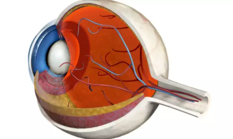
Optic nerve avulsion (ONA) is the traumatic separation of the optic nerve fibers at the level of the lamina cribrosa with preservation of the nerve sheath and surrounding sclera. It is a rare but visually devastating form of anterior traumatic optic neuropathy. ONA can be either partial (only the optic nerve is torn) or complete (both the extraocular muscles and the optic nerve are torn, causing total luxation of the ocular bulb). In cases of complete ONA, the optic sheath, which is more elastic than the optic nerve, usually remains attached to the globe, and the optic nerve may appear normal. A complete avulsion causes profound vision impairment, whereas a partial (incomplete) avulsion might result in varying degrees of impairment.
There are several potential causes of ONA including blunt trauma, road traffic accident (RTA), orautoenucleation in psychiatric patients. ONA has been observed from sports injuries in children, falls, and door handle trauma.
The medical records of patients diagnosed with ONA at an Ophthalmic Emergency Department between November 2014 and November 2022 were analyzed in a retrospective cohort study by Mohammad Al Amry et al. Data were collected on patient age, sex, affected eye, cause of injury and imaging studies. The best-corrected visual acuity (BCVA) at presentation and at the last follow-up visit, and the duration of follow-up were documented.
The study sample was comprised of 44 eyes of 43 patients with ONA with median age of 16.5 (9.3–26.8) years ranging from 2 years old to 70 years old. There were (35; 79.5%) males and (9; 20.5%) females. Most cases presented with an affected left eye (27; 61.4%) followed by the right eye (16; 36.4%) and only one patient (2.3%) had bilateral ONA. The most common cause of trauma resulting in ONA was a metallic object (8; 18.2%). This study demonstrates the value of multi-sequence Magnetic resonance imaging (MRI) in the setting of unexplained vision loss when other modalities are inadequate or inconclusive.
Optic nerve avulsion is a devastating injury. Injuries caused by metallic objects were the leading cause of ONA. The presence of associated media opacities challenges the initial diagnosis of ONA. In the vast majority of cases, the vision ended as NLP, indicating permanent vision impairment.
Source: Mohammad Al Amry, Lamia AlHijji , Sahar M Elkhamary; Clinical Ophthalmology 2023:17 2633–2641
Dr Ishan Kataria has done his MBBS from Medical College Bijapur and MS in Ophthalmology from Dr Vasant Rao Pawar Medical College, Nasik. Post completing MD, he pursuid Anterior Segment Fellowship from Sankara Eye Hospital and worked as a competent phaco and anterior segment consultant surgeon in a trust hospital in Bathinda for 2 years.He is currently pursuing Fellowship in Vitreo-Retina at Dr Sohan Singh Eye hospital Amritsar and is actively involved in various research activities under the guidance of the faculty.
Dr Kamal Kant Kohli-MBBS, DTCD- a chest specialist with more than 30 years of practice and a flair for writing clinical articles, Dr Kamal Kant Kohli joined Medical Dialogues as a Chief Editor of Medical News. Besides writing articles, as an editor, he proofreads and verifies all the medical content published on Medical Dialogues including those coming from journals, studies,medical conferences,guidelines etc. Email: drkohli@medicaldialogues.in. Contact no. 011-43720751


