- Home
- Medical news & Guidelines
- Anesthesiology
- Cardiology and CTVS
- Critical Care
- Dentistry
- Dermatology
- Diabetes and Endocrinology
- ENT
- Gastroenterology
- Medicine
- Nephrology
- Neurology
- Obstretics-Gynaecology
- Oncology
- Ophthalmology
- Orthopaedics
- Pediatrics-Neonatology
- Psychiatry
- Pulmonology
- Radiology
- Surgery
- Urology
- Laboratory Medicine
- Diet
- Nursing
- Paramedical
- Physiotherapy
- Health news
- Fact Check
- Bone Health Fact Check
- Brain Health Fact Check
- Cancer Related Fact Check
- Child Care Fact Check
- Dental and oral health fact check
- Diabetes and metabolic health fact check
- Diet and Nutrition Fact Check
- Eye and ENT Care Fact Check
- Fitness fact check
- Gut health fact check
- Heart health fact check
- Kidney health fact check
- Medical education fact check
- Men's health fact check
- Respiratory fact check
- Skin and hair care fact check
- Vaccine and Immunization fact check
- Women's health fact check
- AYUSH
- State News
- Andaman and Nicobar Islands
- Andhra Pradesh
- Arunachal Pradesh
- Assam
- Bihar
- Chandigarh
- Chattisgarh
- Dadra and Nagar Haveli
- Daman and Diu
- Delhi
- Goa
- Gujarat
- Haryana
- Himachal Pradesh
- Jammu & Kashmir
- Jharkhand
- Karnataka
- Kerala
- Ladakh
- Lakshadweep
- Madhya Pradesh
- Maharashtra
- Manipur
- Meghalaya
- Mizoram
- Nagaland
- Odisha
- Puducherry
- Punjab
- Rajasthan
- Sikkim
- Tamil Nadu
- Telangana
- Tripura
- Uttar Pradesh
- Uttrakhand
- West Bengal
- Medical Education
- Industry
Kneeling Stress Radiography a forgotten yet dependable tool for Posterolateral Knee Instability

Diagnosing postero-lateral knee instability is a challenge from both clinical and radiologic perspective and can lead to significant morbidity if left untreated. Delayed diagnosis leads to a more demanding surgery and prolonged rehabilitation for the patient.
Stress radiography (SR) lost its popularity in recent years due to more dependence on instrumented laxity measurement (KT-1000) along with MRI. Kneeling stress radiograph is a lost art but remains invaluable in the assessment of posterolateral knee instability.
Quamar Azam et al conducted a prospective observational study to re-explore the undeniable utility of this forgotten tool in early diagnosis of posterolateral knee instability and identifying the mean posterior tibial translation distance (PTTD) and also assessing side to side difference (SSD) between the injured and the contralateral normal knee.
All patients with suspected ligament injuries were clinically evaluated using Lachman test, Drawer test (included palpation of tibial step of and posterior sag test, Godfrey test), Pivot shift test, Dial test (at 30° and 90° in the prone position and more than 10° of difference in external rotation from the normal side was considered significant), and Varus-valgus stress tests (at zero and 30°).
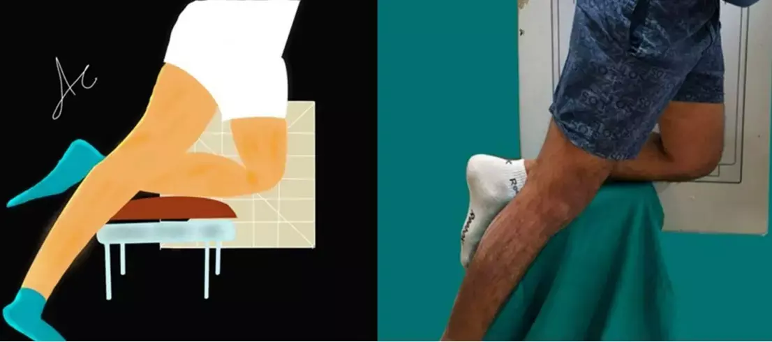
KSR (kneeling stress radiograph) was employed as a routine additional tool besides clinical examination and routine 3 T MRI. Patients were made to stand on one leg while the knee to be examined was flexed to 90° and kept on well-padded but firm support. The thigh was placed perpendicular to the horizontal leg and the patient was asked to rotate approximately 10° towards the examination side to superimpose the femoral condyles. The knee must be as close as possible to the film and the X-ray beam should be perpendicular to the film. Just before acquisition, the patient was asked to bear complete (or near-complete) weight on the knee under examination while the contralateral leg (on the ground) provides proprioception for required balance.
The observations of the study were:
• Total 27 patients were included in the study, with males being 4.4 times more commonly injured as compared to females.
• The most common mode of injury was motor vehicle accident (MVA).
• Out of 27 patients, 11 had isolated PCL (posterior cruciate ligament) injury while the rest had PLC (posterolateral corner) involvement.
• The mean SSD of PTTD was 8.79 mm in patient with isolated PCL. This was increased by 1.65 times (14.52 mm) in patients with co-existing PLC involvement.
The authors commented that – "The three main points to remember include—perpendicular position of knee relative to thigh, both femoral condyles should superimpose, and maximal weight should be born on the knee under examination."
The authors concluded that - Stress radiography is an indelible tool and serves as an adjunct to MRI imaging. It is not a substitute for clinical assessment but assists in early diagnosis and management, facilitating surgical planning and furnishing objective evidence of PCL/PLC functionality which is not possible with any other imaging modality.
Further reading:
Kneeling Stress Radiography: A Forgotten yet Dependable Tool for Postero lateral Knee Instability
Quamar Azam, Abhishek Chandra et al
Indian Journal of Orthopaedics (2022) 56:1729–1736
MBBS, Dip. Ortho, DNB ortho, MNAMS
Dr Supreeth D R (MBBS, Dip. Ortho, DNB ortho, MNAMS) is a practicing orthopedician with interest in medical research and publishing articles. He completed MBBS from mysore medical college, dip ortho from Trivandrum medical college and sec. DNB from Manipal Hospital, Bengaluru. He has expirence of 7years in the field of orthopedics. He has presented scientific papers & posters in various state, national and international conferences. His interest in writing articles lead the way to join medical dialogues. He can be contacted at editorial@medicaldialogues.in.
Dr Kamal Kant Kohli-MBBS, DTCD- a chest specialist with more than 30 years of practice and a flair for writing clinical articles, Dr Kamal Kant Kohli joined Medical Dialogues as a Chief Editor of Medical News. Besides writing articles, as an editor, he proofreads and verifies all the medical content published on Medical Dialogues including those coming from journals, studies,medical conferences,guidelines etc. Email: drkohli@medicaldialogues.in. Contact no. 011-43720751


