Rotational Spine Fractures Linked to High Risk of Acute Cord Injury, Suggests Study
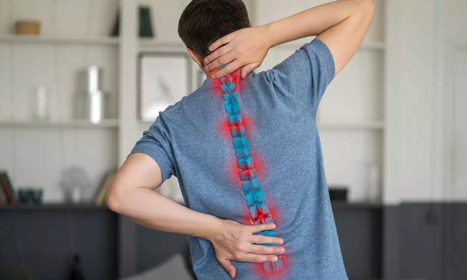


A recent retrospective study published in the Indian Journal of Orthopaedics in September 2026 highlights the severe risk posed by rotational and distraction spine fractures. These high-energy injuries triggered acute spinal cord injury in 50.3% of patients, with cord edema or transection occurring in 45.1%.
Traumatic cervicothoracic spine fractures represent common, devastating injuries that frequently cause acute spinal cord injury (SCI) and persistent neurological disability; while computed tomography (CT) serves as the mainstay for identifying bony fractures, magnetic resonance imaging (MRI) demonstrates enhanced sensitivity for detecting soft-tissue disruption and spinal cord pathology. To bridge the clinical gap regarding how specific skeletal fracture patterns correlate with soft-tissue cord damage, principal author Ali Fakheri and colleagues evaluated the relationship between CT-classified cervicothoracic spine fracture types and acute spinal cord injury parameters visualized on MRI.
Therefore, the single-center retrospective study evaluated 195 trauma patients with cervicothoracic spine injuries who underwent both CT and MRI. Fractures were classified on CT as compression, burst, or rotational/distraction to evaluate their correlation with MRI-detected spinal cord damage.
Key Clinical Findings of the Study Includes:
- Fracture Mechanism Distribution: The study demonstrated that compression fractures and burst fractures each comprised 33.6% of cohort cases, whereas rotational/distraction injuries accounted for 32.8%.
- Spinal Cord Injury Frequency: The study identified acute spinal cord involvement in 50.3% of patients, with cord edema or transection documented in 45.1% and intramedullary hemorrhage observed in 25.1%.
- Associated Soft-Tissue Pathology: The study revealed that epidural hematomas occurred in 43.6% of patients and traumatic intervertebral disk herniations were detected in 17.9% of cases.
- Rotational Mechanism Severity: The study established that MRI evidence of cord compression, cord edema, and intramedullary hemorrhage exhibited a significantly stronger correlation with rotational/distraction fractures than with compression or burst patterns.
The results suggest that rotational and distraction fracture mechanisms carry a substantially elevated risk of causing severe spinal cord damage compared to other injury types, given the overall 50.3% prevalence of acute cord injury. Consequently, early recognition of these specific structural fracture patterns on CT scans is vital for initiating rapid MRI evaluation and neuroprotective strategies.
These findings suggest that practicing clinicians should maintain heightened clinical vigilance for underlying cord parenchymal injury when rotational or distraction fracture configurations are identified on initial computed tomography imaging.
While the single-center retrospective design presents inherent methodological limitations, further prospective multi-center studies could comfortably expand upon these findings to refine diagnostic workflows and risk stratification for acute spinal trauma management.
Reference
Fakheri A, Shakeri A, Motamedi A, Jalili J, Karbasi M, Rezaei S. Relationship Between Different Types of Traumatic Cervicothoracic Spine Fractures on CT Scan with Acute Spinal Cord Injury on MRI. Indian Journal of Orthopaedics. Published online September 4, 2026.