- Home
- Medical news & Guidelines
- Anesthesiology
- Cardiology and CTVS
- Critical Care
- Dentistry
- Dermatology
- Diabetes and Endocrinology
- ENT
- Gastroenterology
- Medicine
- Nephrology
- Neurology
- Obstretics-Gynaecology
- Oncology
- Ophthalmology
- Orthopaedics
- Pediatrics-Neonatology
- Psychiatry
- Pulmonology
- Radiology
- Surgery
- Urology
- Laboratory Medicine
- Diet
- Nursing
- Paramedical
- Physiotherapy
- Health news
- Fact Check
- Bone Health Fact Check
- Brain Health Fact Check
- Cancer Related Fact Check
- Child Care Fact Check
- Dental and oral health fact check
- Diabetes and metabolic health fact check
- Diet and Nutrition Fact Check
- Eye and ENT Care Fact Check
- Fitness fact check
- Gut health fact check
- Heart health fact check
- Kidney health fact check
- Medical education fact check
- Men's health fact check
- Respiratory fact check
- Skin and hair care fact check
- Vaccine and Immunization fact check
- Women's health fact check
- AYUSH
- State News
- Andaman and Nicobar Islands
- Andhra Pradesh
- Arunachal Pradesh
- Assam
- Bihar
- Chandigarh
- Chattisgarh
- Dadra and Nagar Haveli
- Daman and Diu
- Delhi
- Goa
- Gujarat
- Haryana
- Himachal Pradesh
- Jammu & Kashmir
- Jharkhand
- Karnataka
- Kerala
- Ladakh
- Lakshadweep
- Madhya Pradesh
- Maharashtra
- Manipur
- Meghalaya
- Mizoram
- Nagaland
- Odisha
- Puducherry
- Punjab
- Rajasthan
- Sikkim
- Tamil Nadu
- Telangana
- Tripura
- Uttar Pradesh
- Uttrakhand
- West Bengal
- Medical Education
- Industry
Traction MRI examination useful for evaluating cartilage lesions at the knee joint

Lesions of the articular cartilage of the knee, especially early grades, are not always accurately detected by magnetic resonance imaging (MRI) because of contact between the articular cartilage surfaces of the femur and the tibia.
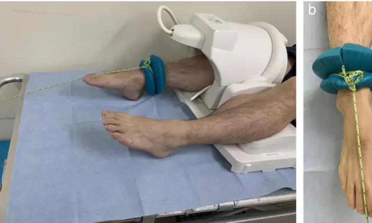
Naoya Kikuchi et al conducted a study to assess the effects of axial leg traction during knee MRI examination on joint space widening and articular cartilage visualization and evaluate the ideal weight for traction. They found that - traction MRI examination may be useful in evaluating articular cartilage lesions at the medial tibiofemoral joint. A traction weight of 5 kg may be sufficient with minimum pain and discomfort. The study has been published in 'Skeletal Radiology' journal.
MRI was performed on ten healthy volunteers using a 3-T MRI unit with a 3D dual-echo steady-state gradientrecalled echo sequence. Conventional MRI was performed first, followed by traction MRI. The traction weight increased in the order of 5 kg, 10 kg, and 15 kg. Joint space widths were measured, and articular cartilage visualization was assessed at the medial and lateral tibiofemoral joints. Volunteers were asked to evaluate pain and discomfort using a visual analog scale during each procedure with axial traction to assess the safety of traction MRI.
The observations in the study were :
• The medial tibiofemoral joint space width significantly increased, and the visualization of the articular cartilage significantly improved by applying traction.
• The joint space width and the articular cartilage visualization showed no significant differences among traction weights of 5 kg, 10 kg, and 15 kg.
{The joint space width at the medial tibiofemoral joint MRI and traction MRI with weights of 5 kg, 10 kg, and 15 kg was 0.5 ± 0.5 mm and 1.8 ± 0.7 mm, 2.1 ± 0.8 mm, and 2.1 ± 0.6 mm respectively (p< 0.00009). Post hoc analysis showed significant differences between MRI and traction MRI with each traction weight (traction weights of 5 kg, 10 kg, and 15 kg; p = 0.028, 0.028, and 0.028, respectively). There were no significant differences between traction weights of 5 kg and 10 kg (p = 0.83), 5 kg and 15 kg (p=0.44), and 10 kg and 15 kg (p=1.00). }
• In contrast, the joint space width at the lateral tibiofemoral joint did not increase significantly with traction (p=0.86)
• Pain and discomfort during traction MRI examination were lowest with a traction weight of 5 kg.
The authors concluded that – their study suggests that traction MRI with a traction weight of 5 kg is possibly effective enough to investigate articular cartilage lesions at the medial tibiofemoral joint.
Further reading:
Improving visualization of the articular cartilage of the knee with magnetic resonance imaging under axial traction: a comparative study of different traction weights
Naoya Kikuchi, Sho Kohyama et al
Skeletal Radiology (2022) 51:1483–1491
https://doi.org/10.1007/s00256-021-03971-w
MBBS, Dip. Ortho, DNB ortho, MNAMS
Dr Supreeth D R (MBBS, Dip. Ortho, DNB ortho, MNAMS) is a practicing orthopedician with interest in medical research and publishing articles. He completed MBBS from mysore medical college, dip ortho from Trivandrum medical college and sec. DNB from Manipal Hospital, Bengaluru. He has expirence of 7years in the field of orthopedics. He has presented scientific papers & posters in various state, national and international conferences. His interest in writing articles lead the way to join medical dialogues. He can be contacted at editorial@medicaldialogues.in.
Dr Kamal Kant Kohli-MBBS, DTCD- a chest specialist with more than 30 years of practice and a flair for writing clinical articles, Dr Kamal Kant Kohli joined Medical Dialogues as a Chief Editor of Medical News. Besides writing articles, as an editor, he proofreads and verifies all the medical content published on Medical Dialogues including those coming from journals, studies,medical conferences,guidelines etc. Email: drkohli@medicaldialogues.in. Contact no. 011-43720751


