- Home
- Medical news & Guidelines
- Anesthesiology
- Cardiology and CTVS
- Critical Care
- Dentistry
- Dermatology
- Diabetes and Endocrinology
- ENT
- Gastroenterology
- Medicine
- Nephrology
- Neurology
- Obstretics-Gynaecology
- Oncology
- Ophthalmology
- Orthopaedics
- Pediatrics-Neonatology
- Psychiatry
- Pulmonology
- Radiology
- Surgery
- Urology
- Laboratory Medicine
- Diet
- Nursing
- Paramedical
- Physiotherapy
- Health news
- Fact Check
- Bone Health Fact Check
- Brain Health Fact Check
- Cancer Related Fact Check
- Child Care Fact Check
- Dental and oral health fact check
- Diabetes and metabolic health fact check
- Diet and Nutrition Fact Check
- Eye and ENT Care Fact Check
- Fitness fact check
- Gut health fact check
- Heart health fact check
- Kidney health fact check
- Medical education fact check
- Men's health fact check
- Respiratory fact check
- Skin and hair care fact check
- Vaccine and Immunization fact check
- Women's health fact check
- AYUSH
- State News
- Andaman and Nicobar Islands
- Andhra Pradesh
- Arunachal Pradesh
- Assam
- Bihar
- Chandigarh
- Chattisgarh
- Dadra and Nagar Haveli
- Daman and Diu
- Delhi
- Goa
- Gujarat
- Haryana
- Himachal Pradesh
- Jammu & Kashmir
- Jharkhand
- Karnataka
- Kerala
- Ladakh
- Lakshadweep
- Madhya Pradesh
- Maharashtra
- Manipur
- Meghalaya
- Mizoram
- Nagaland
- Odisha
- Puducherry
- Punjab
- Rajasthan
- Sikkim
- Tamil Nadu
- Telangana
- Tripura
- Uttar Pradesh
- Uttrakhand
- West Bengal
- Medical Education
- Industry
Novel Modification of Extended Trochanteric Osteotomy may save greater trochanter

The extended trochanteric osteotomy is the workhorse for removal of well-fixed femoral stems during total hip revision arthroplasty. Despite its reliable performance in exposing the implants for removal and accessing the femoral canal, significant complications can occur. Though these complications are rare, trochanteric nonunion, trochanteric escape, and femoral implant subsidence can have a significant negative impact on gait mechanics and patient outcome. If access to the canal was still possible and the greater trochanter could remain in place, these complications could be minimized or possibly even eliminated.
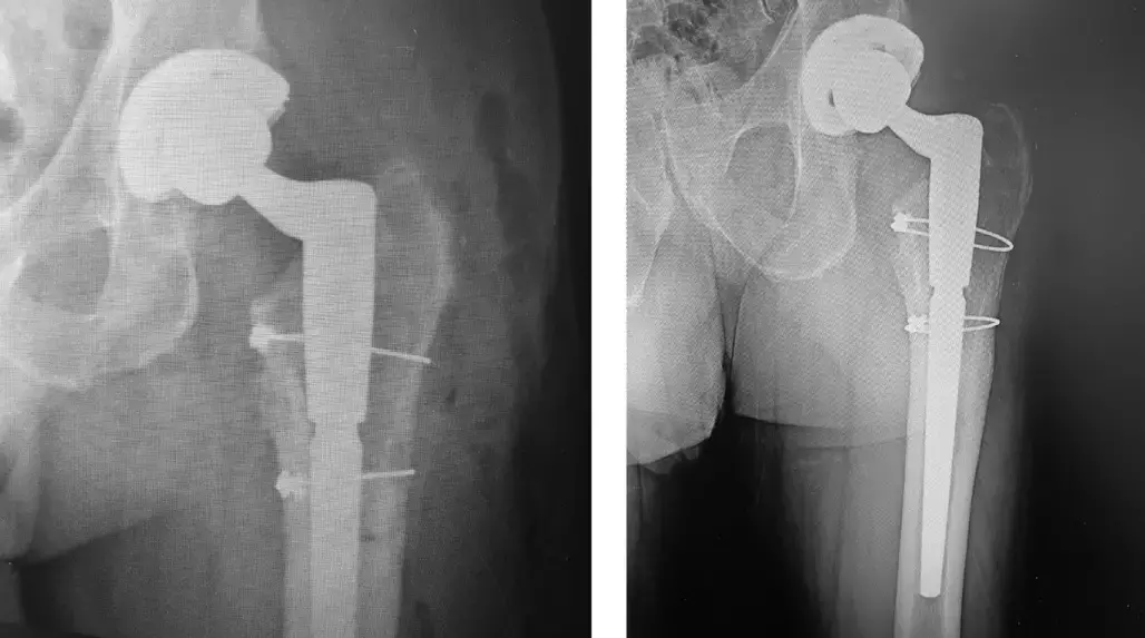
The paper by Eric B. Smith has been published in 'arthroplasty today' journal. It describes a novel technique using a lateral cortical window just distal to the greater trochanter that allows removal of a well-fixed stem and leaves the greater trochanter intact.
This case report describes the procedure on a 77-year-old male who underwent his primary THA 10 years prior to the revision. He presented with sudden onset of severe groin pain and difficulty ambulating. Radiographs revealed the cementless stem had catastrophic failure of the trunnion with complete dissociation of the cobalt chromium femoral head.
Though the patient never complained of pain previously, radiographs of the femur showed lucencies at zone 1 and 7, lack of calcar atrophy, and pedestal formation at the tip of the stem. The acetabulum appeared to be well fixed, and revision of the femoral stem was planned.
This surgery was performed via a direct lateral approach; however, any approach that can access the proximal and lateral aspect of the femur can be utilized. The basic preparation is the same as the ETO as described by MacDonald et al. After dislocation of the hip and removal of the femoral head, the proximal femur was cleared of debris to visualize the entire proximal bone implant interface. A 2-mm burr and flexible osteotomes were used to remove any points of fixation proximally between the implant and the bone. As these devices are passed distally, great care is necessary to ensure no perforation of the cortical bone. An extraction device was placed on the stem, yet the implant would not release. It was decided to perform the osteotomy.
The borders of the osteotomy were identified. The proximal aspect is just distal to vastus ridge ensuring 2 cm of bone is not disrupted proximally. The distal aspect was measured 6 cm distal to the proximal cut- this was determined based on the porous coating of the stem. It is important that the osteotomy extends beyond the porous coating of the stem so as to allow for release of distal spot welds into the stem. The anterior cut was made at the lateral edge of the implant, and the posterior cut was made internally after the anterior cut was made. The anterior cut was made with the oscillating saw. The proximal and distal cuts were made with a pencil tip burr, and the posterior cut was made through the anterior cut to score the inner cortex. Osteotomes were used to lever out the fragment, leaving the greater trochanter completely intact. This was performed similarly to the standard ETO, but the proximal extent was stopped at the vastus ridge. The osteotomized fragment was levered posteriorly, and the stem was visualized.
The flexible osteotome was passed through the osteotomy, proximally ensuring the Gigli could be passed through without obstruction. The Gigli saw was passed proximally through the osteotomy and around the implant and then used in a sawing fashion moving distally until it passes the distal aspect of the porous coating. The extraction handle was attached to the implant, and the stem was removed with ease and without any appreciable bone loss. The osteotomy fragment was then secured in anatomic position with 2 cables.
The femur was then prepared in routine fashion with a modular stem. The wound was closed in routine fashion. Postoperative radiographs show anatomic position of the osteotomized bone fragment and the greater trochanter intact. The patient performed 50% weight-bearing for the first 4 weeks and then advanced as tolerated. This limitation was more for the style of femoral implant than for protection of the osteotomized fragment. He progressed without difficulty and ambulated without a limp, and his radiographs at 1 year revealed complete healing of the osteotomy and no stem subsidence.
Further reading:
Save the Greater Trochanter: A Novel Modification to the Extended Trochanteric Osteotomy Eric B. Smith Arthroplasty Today 16 (2022) 107- 111 https://doi.org/10.1016/j.artd.2022.05.004
MBBS, Dip. Ortho, DNB ortho, MNAMS
Dr Supreeth D R (MBBS, Dip. Ortho, DNB ortho, MNAMS) is a practicing orthopedician with interest in medical research and publishing articles. He completed MBBS from mysore medical college, dip ortho from Trivandrum medical college and sec. DNB from Manipal Hospital, Bengaluru. He has expirence of 7years in the field of orthopedics. He has presented scientific papers & posters in various state, national and international conferences. His interest in writing articles lead the way to join medical dialogues. He can be contacted at editorial@medicaldialogues.in.
Dr Kamal Kant Kohli-MBBS, DTCD- a chest specialist with more than 30 years of practice and a flair for writing clinical articles, Dr Kamal Kant Kohli joined Medical Dialogues as a Chief Editor of Medical News. Besides writing articles, as an editor, he proofreads and verifies all the medical content published on Medical Dialogues including those coming from journals, studies,medical conferences,guidelines etc. Email: drkohli@medicaldialogues.in. Contact no. 011-43720751


