- Home
- Medical news & Guidelines
- Anesthesiology
- Cardiology and CTVS
- Critical Care
- Dentistry
- Dermatology
- Diabetes and Endocrinology
- ENT
- Gastroenterology
- Medicine
- Nephrology
- Neurology
- Obstretics-Gynaecology
- Oncology
- Ophthalmology
- Orthopaedics
- Pediatrics-Neonatology
- Psychiatry
- Pulmonology
- Radiology
- Surgery
- Urology
- Laboratory Medicine
- Diet
- Nursing
- Paramedical
- Physiotherapy
- Health news
- Fact Check
- Bone Health Fact Check
- Brain Health Fact Check
- Cancer Related Fact Check
- Child Care Fact Check
- Dental and oral health fact check
- Diabetes and metabolic health fact check
- Diet and Nutrition Fact Check
- Eye and ENT Care Fact Check
- Fitness fact check
- Gut health fact check
- Heart health fact check
- Kidney health fact check
- Medical education fact check
- Men's health fact check
- Respiratory fact check
- Skin and hair care fact check
- Vaccine and Immunization fact check
- Women's health fact check
- AYUSH
- State News
- Andaman and Nicobar Islands
- Andhra Pradesh
- Arunachal Pradesh
- Assam
- Bihar
- Chandigarh
- Chattisgarh
- Dadra and Nagar Haveli
- Daman and Diu
- Delhi
- Goa
- Gujarat
- Haryana
- Himachal Pradesh
- Jammu & Kashmir
- Jharkhand
- Karnataka
- Kerala
- Ladakh
- Lakshadweep
- Madhya Pradesh
- Maharashtra
- Manipur
- Meghalaya
- Mizoram
- Nagaland
- Odisha
- Puducherry
- Punjab
- Rajasthan
- Sikkim
- Tamil Nadu
- Telangana
- Tripura
- Uttar Pradesh
- Uttrakhand
- West Bengal
- Medical Education
- Industry
18F-FDG PET/CT bests ultrasound for early detection of lymph node and abdominal metastases in melanoma

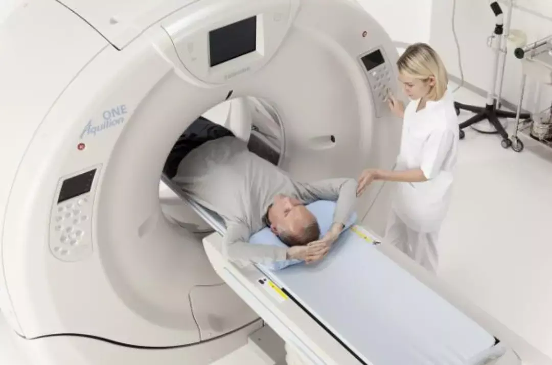
According to a new study 2-18fluoro-2-deoxy-D-glucose positron emission tomography/computed tomography (18F-FDG PET/CT) is preferred over Ultrsound for early detection of lymph node and abdominal metastases among patients with malignant melanoma.
The study has been published in Medicine Journal.
Malignant melanoma (MM) has become more common over time. According to the TNM system, which describes the size of the tumor (T), the number of infected lymph nodes (N), and the existence of organ metastases (M), MM is categorized. Patients with MM get either 18F-FDG PET/CT or ultrasonography (US) of the abdomen (aUS) and peripheral lymph nodes (pUS) (cervical, axillary, and inguinal). This study was carried out by Philipp Weber and colleagues to assess the sensitivity and specificity of 18F-FDG PET/CT and ultrasonography for staging malignant melanoma patients.
There were 258 patients in all who satisfied the primary inclusion criteria for malignant melanoma without additional cancer that was shown by histology (mean age, 61 16 years; 112 men and 146 women). This analysis of the diagnostic precision was retrospective. The patient and radiological information system at the hospital served as the source of all data. Patients were investigated by 18F-FDG PET/CT and 176 more times by US, for a total of 584 18F-FDG PET/CT and 697 extra times. Both the US and 18F-FDG PET/CT showed 824 and 726 lesions, respectively. Additionally, analysis by patient, examination, and lesion were carried out. Histopathology, excision of lesions, and follow-up controls utilizing additional imaging techniques served as the reference standards.
The key findings of this study were:
1. Both the sensitivity of 18F-FDG PET/CT (0.80) comparing to wUS (0.63) and pUS (0.61) and the precision of 18F-FDG PET/CT (0.96) compared to wUS (0.98) and aUS were found to be significantly different (P .05) in the per-examination (0.99).
2. In the PLA, there were significant differences between wUS (0.61, 0.98), pUS (0.60, 0.98), and aUS in terms of sensitivity and specificity for 18F-FDG PET/CT (0.83, 0.91). (0.61, 0.99).
In comparison to the US, 18F-FDG PET/CT has a somewhat lower specificity but a better sensitivity. When compared to the sensitivity of 18F-FDG PET/CT alone, the combined use of 18F-FDG PET/CT and US shows a modest benefit. The solitary use of 18F-FDG PET/CT and US, as well as the combined use of these 2 radiological modalities, are equal in terms of specificity. Overall, when expenses are taken into account, using 18F-FDG PET/CT as the only application is preferred. Only a few benefits of combined examination may be cited.
Reference:
Weber, P., Arnold, A., & Hohmann, J. (2022). Comparison of 18F-FDG PET/CT and ultrasound in staging of patients with malignant melanoma. In Medicine (Vol. 101, Issue 42, p. e31092). Ovid Technologies (Wolters Kluwer Health). https://doi.org/10.1097/md.0000000000031092
Neuroscience Masters graduate
Jacinthlyn Sylvia, a Neuroscience Master's graduate from Chennai has worked extensively in deciphering the neurobiology of cognition and motor control in aging. She also has spread-out exposure to Neurosurgery from her Bachelor’s. She is currently involved in active Neuro-Oncology research. She is an upcoming neuroscientist with a fiery passion for writing. Her news cover at Medical Dialogues feature recent discoveries and updates from the healthcare and biomedical research fields. She can be reached at editorial@medicaldialogues.in
Dr Kamal Kant Kohli-MBBS, DTCD- a chest specialist with more than 30 years of practice and a flair for writing clinical articles, Dr Kamal Kant Kohli joined Medical Dialogues as a Chief Editor of Medical News. Besides writing articles, as an editor, he proofreads and verifies all the medical content published on Medical Dialogues including those coming from journals, studies,medical conferences,guidelines etc. Email: drkohli@medicaldialogues.in. Contact no. 011-43720751


