- Home
- Medical news & Guidelines
- Anesthesiology
- Cardiology and CTVS
- Critical Care
- Dentistry
- Dermatology
- Diabetes and Endocrinology
- ENT
- Gastroenterology
- Medicine
- Nephrology
- Neurology
- Obstretics-Gynaecology
- Oncology
- Ophthalmology
- Orthopaedics
- Pediatrics-Neonatology
- Psychiatry
- Pulmonology
- Radiology
- Surgery
- Urology
- Laboratory Medicine
- Diet
- Nursing
- Paramedical
- Physiotherapy
- Health news
- Fact Check
- Bone Health Fact Check
- Brain Health Fact Check
- Cancer Related Fact Check
- Child Care Fact Check
- Dental and oral health fact check
- Diabetes and metabolic health fact check
- Diet and Nutrition Fact Check
- Eye and ENT Care Fact Check
- Fitness fact check
- Gut health fact check
- Heart health fact check
- Kidney health fact check
- Medical education fact check
- Men's health fact check
- Respiratory fact check
- Skin and hair care fact check
- Vaccine and Immunization fact check
- Women's health fact check
- AYUSH
- State News
- Andaman and Nicobar Islands
- Andhra Pradesh
- Arunachal Pradesh
- Assam
- Bihar
- Chandigarh
- Chattisgarh
- Dadra and Nagar Haveli
- Daman and Diu
- Delhi
- Goa
- Gujarat
- Haryana
- Himachal Pradesh
- Jammu & Kashmir
- Jharkhand
- Karnataka
- Kerala
- Ladakh
- Lakshadweep
- Madhya Pradesh
- Maharashtra
- Manipur
- Meghalaya
- Mizoram
- Nagaland
- Odisha
- Puducherry
- Punjab
- Rajasthan
- Sikkim
- Tamil Nadu
- Telangana
- Tripura
- Uttar Pradesh
- Uttrakhand
- West Bengal
- Medical Education
- Industry
Dark-field chest X-ray helpful for differentiating emphysema from fibrosis: Study

Germany: Researchers from Germany in a recent case study published in Radiology: Cardiothoracic Imaging have discussed a prototype dark-field chest x-ray system that may be used for differentiating between fibrosis and emphysema.
The case described by Florian T. Gassert and colleagues is of an 83-year-old man who presented at their hospital with combined pulmonary fibrosis and emphysema (CPFE). Chest CT scan showed advanced upper lung mixed centrilobular and paraseptal emphysema and lower lung–predominant peripheral fibrotic changes with honeycombing, indicating a definite usual interstitial pneumonia pattern. Pulmonary function tests showed combined obstructive to forced vital capacity and restrictive lung changes, with impaired lung diffusion capacity.
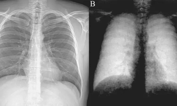
The researchers used the prototype system that produces both a dark-field chest radiography (dfCXR) dfCXR and a conventional attenuation-based radiograph (atCXR) in a single scan.
The patient demonstrated:
- Substantially decreased dark-field signal in the upper lungs (left more than right), where emphysema is most severe, and inhomogeneous but decreased lower lung signal (due to presence of fibrosis) compared to a healthy subject.
- The stronger manifestation of fibrosis in the right lower lobe compared with the left lower lobe, as was seen on CT images, corresponded well to the asymmetric reduction of dark-field signal in the lower fields with a right-sided predominance.
The researchers wrote, "as both fibrosis and emphysema result in a reduction of dark-field signal–due to a smaller number of intact alveoli–the assignment of signal loss to one of the two pathologic conditions is hardly possible from the dfCXR alone. However, the combo of atCXR and dfCXR might allow for the differentiation of emphysema and fibrosis, particularly as images are perfectly co-registered, they suggested.
"While there is a need for the further investigation of dark-field signal appearance of fibrosis, first studies have shown that dfCXR allows for the qualitative and quantitative assessment of emphysema, depicting regional and global lung impairment," they concluded.
Reference:
The study titled, "Dark-Field Chest Radiography of Combined Pulmonary Fibrosis and Emphysema," was published in Radiology: Cardiothoracic Imaging.
DOI: https://doi.org/10.1148/ryct.220085
Dr Kamal Kant Kohli-MBBS, DTCD- a chest specialist with more than 30 years of practice and a flair for writing clinical articles, Dr Kamal Kant Kohli joined Medical Dialogues as a Chief Editor of Medical News. Besides writing articles, as an editor, he proofreads and verifies all the medical content published on Medical Dialogues including those coming from journals, studies,medical conferences,guidelines etc. Email: drkohli@medicaldialogues.in. Contact no. 011-43720751


