- Home
- Medical news & Guidelines
- Anesthesiology
- Cardiology and CTVS
- Critical Care
- Dentistry
- Dermatology
- Diabetes and Endocrinology
- ENT
- Gastroenterology
- Medicine
- Nephrology
- Neurology
- Obstretics-Gynaecology
- Oncology
- Ophthalmology
- Orthopaedics
- Pediatrics-Neonatology
- Psychiatry
- Pulmonology
- Radiology
- Surgery
- Urology
- Laboratory Medicine
- Diet
- Nursing
- Paramedical
- Physiotherapy
- Health news
- Fact Check
- Bone Health Fact Check
- Brain Health Fact Check
- Cancer Related Fact Check
- Child Care Fact Check
- Dental and oral health fact check
- Diabetes and metabolic health fact check
- Diet and Nutrition Fact Check
- Eye and ENT Care Fact Check
- Fitness fact check
- Gut health fact check
- Heart health fact check
- Kidney health fact check
- Medical education fact check
- Men's health fact check
- Respiratory fact check
- Skin and hair care fact check
- Vaccine and Immunization fact check
- Women's health fact check
- AYUSH
- State News
- Andaman and Nicobar Islands
- Andhra Pradesh
- Arunachal Pradesh
- Assam
- Bihar
- Chandigarh
- Chattisgarh
- Dadra and Nagar Haveli
- Daman and Diu
- Delhi
- Goa
- Gujarat
- Haryana
- Himachal Pradesh
- Jammu & Kashmir
- Jharkhand
- Karnataka
- Kerala
- Ladakh
- Lakshadweep
- Madhya Pradesh
- Maharashtra
- Manipur
- Meghalaya
- Mizoram
- Nagaland
- Odisha
- Puducherry
- Punjab
- Rajasthan
- Sikkim
- Tamil Nadu
- Telangana
- Tripura
- Uttar Pradesh
- Uttrakhand
- West Bengal
- Medical Education
- Industry
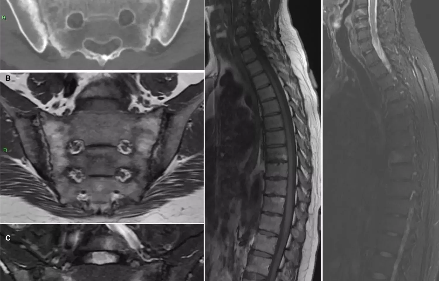
Findings on spine MRI may not always indicate spondyloarthritis: Study

Belgium: Progressive increase in sacroiliac joint and spinal lesions detected on MRI are frequently seen in healthy individuals, especially in older subjects and are not always the indicator of spondyloarthritis, reports a study published in the Arthritis & Rheumatology.
Magnetic Resonance Imaging (MRI) is the most sensitive imaging technique to detect sacroiliitis, a primary manifestation of axial spondyloarthritis (AxSpA). Bone marrow oedema (BMO) on MRI of sacroiliac joints (SIJs) represents a hallmark of axial spondyloarthritis (SpA), yet studies have shown that such lesions may also occur under augmented mechanical stress in healthy subjects, such as postpartum women, military recruits, and young athletes. MRI plays a pivotal role in spondyloarthritis (SpA) diagnosis. However, a detailed description of MRI findings of the sacroiliac (SI) joints and spine in healthy individuals is currently lacking.
The present study was conducted by Thomas Renson, Ghent University, Ghent, Belgium and his team to evaluate the occurrence of MRI-detected SI joint and spinal lesions in healthy individuals in relation to age.
Researchers included 95 healthy subjects (ages 20–49 years) in the study who underwent an MRI of the SI joints and spine. Bone marrow oedema (BME) and structural lesions of the SI joints were scored using the Spondyloarthritis Research Consortium of Canada (SPARCC) method. Spinal inflammatory and structural lesions were evaluated using the SPARCC MRI spine inflammation index and the Canada-Denmark MRI scoring system, respectively. The research team reviewed the fulfilment of the Assessment of SpondyloArthritis international Society's definition of a positive MRI for sacroiliitis/spondylitis. Findings were compared to MRIs of axial SpA patients from the Belgian Inflammatory Arthritis and Spondylitis cohort.
Key findings of the study,
• 17.2% of the subjects ≥30 years old, fulfilled the definition of a positive MRI for sacroiliitis, but this was rarely observed in younger subjects.
• SI joint erosions (20.0%) and fat metaplasia (13.7%) were detected across all age groups.
• Erosions were more frequently visualized in subjects ages ≥40 years (39.3%).
• Spinal BME (35.7%) and fat metaplasia (28.6%) were common in subjects older than 40 years
• Only 1 subject had ≥3 corner inflammatory lesions, indicative of an autoimmune process.
• SI joint and spinal SPARCC scores and total structural lesions scores increased progressively with age.
The authors conclude that contrary to the common belief, structural MRI-detected SI joint lesions can be frequently seen in healthy individuals. Especially in older subjects, the high occurrence of inflammatory and structural MRI-detected lesions impacts their specificity for SpA, which has important implications for the interpretation of MRIs in patients with a clinical suspicion of SpA.
Based on present study data, the authors suggest, "rheumatologists should be vigilant while diagnosing spondyloarthritis (SpA) based on imaging.
Reference:
Renson T, de Hooge M, De Craemer AS, Deroo L, Lukasik Z, Carron P, Herregods N, Jans L, Colman R, Van den Bosch F, Elewaut D. Progressive Increase in Sacroiliac Joint and Spinal Lesions Detected on Magnetic Resonance Imaging in Healthy Individuals in Relation to Age. Arthritis Rheumatol. 2022 Apr 18. doi: 10.1002/art.42145.
BDS
Dr. Hiral patel (BDS) has completed BDS from Gujarat University, Baroda. She has worked in private dental steup for 8years and is currently a consulting general dentist in mumbai. She has recently completed her advanced PG diploma in clinical research and pharmacovigilance. She is passionate about writing and loves to read, analyses and write informative medical content for readers. She can be contacted at editorial@medicaldialogues.in.
Dr Kamal Kant Kohli-MBBS, DTCD- a chest specialist with more than 30 years of practice and a flair for writing clinical articles, Dr Kamal Kant Kohli joined Medical Dialogues as a Chief Editor of Medical News. Besides writing articles, as an editor, he proofreads and verifies all the medical content published on Medical Dialogues including those coming from journals, studies,medical conferences,guidelines etc. Email: drkohli@medicaldialogues.in. Contact no. 011-43720751


