- Home
- Medical news & Guidelines
- Anesthesiology
- Cardiology and CTVS
- Critical Care
- Dentistry
- Dermatology
- Diabetes and Endocrinology
- ENT
- Gastroenterology
- Medicine
- Nephrology
- Neurology
- Obstretics-Gynaecology
- Oncology
- Ophthalmology
- Orthopaedics
- Pediatrics-Neonatology
- Psychiatry
- Pulmonology
- Radiology
- Surgery
- Urology
- Laboratory Medicine
- Diet
- Nursing
- Paramedical
- Physiotherapy
- Health news
- Fact Check
- Bone Health Fact Check
- Brain Health Fact Check
- Cancer Related Fact Check
- Child Care Fact Check
- Dental and oral health fact check
- Diabetes and metabolic health fact check
- Diet and Nutrition Fact Check
- Eye and ENT Care Fact Check
- Fitness fact check
- Gut health fact check
- Heart health fact check
- Kidney health fact check
- Medical education fact check
- Men's health fact check
- Respiratory fact check
- Skin and hair care fact check
- Vaccine and Immunization fact check
- Women's health fact check
- AYUSH
- State News
- Andaman and Nicobar Islands
- Andhra Pradesh
- Arunachal Pradesh
- Assam
- Bihar
- Chandigarh
- Chattisgarh
- Dadra and Nagar Haveli
- Daman and Diu
- Delhi
- Goa
- Gujarat
- Haryana
- Himachal Pradesh
- Jammu & Kashmir
- Jharkhand
- Karnataka
- Kerala
- Ladakh
- Lakshadweep
- Madhya Pradesh
- Maharashtra
- Manipur
- Meghalaya
- Mizoram
- Nagaland
- Odisha
- Puducherry
- Punjab
- Rajasthan
- Sikkim
- Tamil Nadu
- Telangana
- Tripura
- Uttar Pradesh
- Uttrakhand
- West Bengal
- Medical Education
- Industry
PET/CT valuable for distinguishing benign from malignant portal vein thrombosis in liver cirrhosis

Egypt: A small retrospective study has revealed that 18F-FDG PET/CT could be a valuable new imaging technique in differentiating between benign and malignant portal vein thrombosis (PVT) in patients with liver cirrhosis.
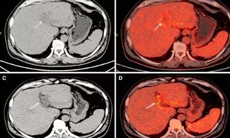
The study, published in the Egyptian Journal of Radiology and Nuclear Medicine, showed that FDG radiotracer uptake on patient F-18 FDG-PET/CT scans was greater when portal vein thrombi were tumours than benign ("bland") thrombi.
"Tumour thrombosis diagnosis and its differentiation from benign thrombosis are critical for managing patients, planning treatments, and minimizing unneeded anticoagulation therapy," Sameh Abokoura, Menoufia University, Shebin Elkoum, Menoufia, Egypt, and colleagues wrote in their study.
Portal vein thrombosis is the complete or partial obstruction of blood flow in the portal vein to the liver due to a thrombus presence. Bland thrombi occur in cancer and non-cancer patients; tumour thrombi and bland can coexist. 18F-FDG PET/CT (18F-fluorodeoxyglucose positron emission tomography/computed tomography) is useful in detecting and diagnosing thrombosis and differentiating it from benign thrombosis.
Abokoura and colleagues aimed to evaluate the value of 18F-FDG PET/CT in distinguishing benign from malignant portal vein thrombosis in patients with liver cirrhosis.
For this purpose, the researchers analyzed 38 patients with liver cirrhosis and portal vein thrombosis, either with or without malignant hepatic tumours, who underwent F-18 FDG-PET/CT scans at their hospital in Menoufia between 2021 and 2022. The semiqualitative analysis included data on all patients' FDG tracer maximum standard uptake (SUVmax) values.
The authors reported the following findings:
- The SUVmax values were significantly higher in the tumour thrombosis group (6.26 ± 1.94) compared to the bland thrombosis group (1.79 ± 0.69).
- The ROC curve of semiqualitative analysis (SUVmax) revealed a specificity of 36.4% and sensitivity of 96.3% at the area under curve of 0.827 with SUVmax > 3.5 as the pathological cut-off value to distinguish tumour from bland thrombi.
The group noted while portal vein thrombosis is frequently associated with liver cirrhosis, it may also occur in patients with cancer, pancreatitis, abdominal sepsis, systemic lupus erythematosus, or other conditions involving hypercoagulable states.
"using semiqualitative analysis, 18F-FDG PET/CT is a valuable new technique in differentiating neoplastic from bland PV thrombi, with optimal cut-off SUVmax value > 3.5 as a criterion," Abokoura and the team concluded.
The limitation of the study was that in 7 cases, imaging and clinical findings were used first to determine the thrombosis type rather than histopathological evidence. Also, the study had flaws due to the retrospective study design and the relatively small patients' number included.
Reference:
Abokoura, S., Ellaban, H. S., & Aly, R. A. (2023). The role of 18F-FDG PET/CT in distinguishing benign from malignant portal vein thrombosis. Egyptian Journal of Radiology and Nuclear Medicine, 54(1), 1-10. https://doi.org/10.1186/s43055-023-01058-1
Dr Kamal Kant Kohli-MBBS, DTCD- a chest specialist with more than 30 years of practice and a flair for writing clinical articles, Dr Kamal Kant Kohli joined Medical Dialogues as a Chief Editor of Medical News. Besides writing articles, as an editor, he proofreads and verifies all the medical content published on Medical Dialogues including those coming from journals, studies,medical conferences,guidelines etc. Email: drkohli@medicaldialogues.in. Contact no. 011-43720751


