- Home
- Medical news & Guidelines
- Anesthesiology
- Cardiology and CTVS
- Critical Care
- Dentistry
- Dermatology
- Diabetes and Endocrinology
- ENT
- Gastroenterology
- Medicine
- Nephrology
- Neurology
- Obstretics-Gynaecology
- Oncology
- Ophthalmology
- Orthopaedics
- Pediatrics-Neonatology
- Psychiatry
- Pulmonology
- Radiology
- Surgery
- Urology
- Laboratory Medicine
- Diet
- Nursing
- Paramedical
- Physiotherapy
- Health news
- Fact Check
- Bone Health Fact Check
- Brain Health Fact Check
- Cancer Related Fact Check
- Child Care Fact Check
- Dental and oral health fact check
- Diabetes and metabolic health fact check
- Diet and Nutrition Fact Check
- Eye and ENT Care Fact Check
- Fitness fact check
- Gut health fact check
- Heart health fact check
- Kidney health fact check
- Medical education fact check
- Men's health fact check
- Respiratory fact check
- Skin and hair care fact check
- Vaccine and Immunization fact check
- Women's health fact check
- AYUSH
- State News
- Andaman and Nicobar Islands
- Andhra Pradesh
- Arunachal Pradesh
- Assam
- Bihar
- Chandigarh
- Chattisgarh
- Dadra and Nagar Haveli
- Daman and Diu
- Delhi
- Goa
- Gujarat
- Haryana
- Himachal Pradesh
- Jammu & Kashmir
- Jharkhand
- Karnataka
- Kerala
- Ladakh
- Lakshadweep
- Madhya Pradesh
- Maharashtra
- Manipur
- Meghalaya
- Mizoram
- Nagaland
- Odisha
- Puducherry
- Punjab
- Rajasthan
- Sikkim
- Tamil Nadu
- Telangana
- Tripura
- Uttar Pradesh
- Uttrakhand
- West Bengal
- Medical Education
- Industry
Breast MRI identifies lesions in patients with normal mammogram, ultrasound: Study

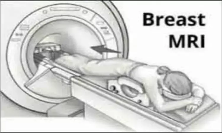
France: Breast MRI can be useful for the management of patients with suspicious nipple discharge and normal ultrasound and mammogram, finds a recent study in the journal European Radiology. In such patients, MRI demonstrated excellent performance for the identification of lesions that required excision. Normal MRI indicates it is safe to propose follow-up only management, thus avoiding unnecessary duct excision.
Martine Boisserie-Lacroix, Department of Radiology, Institut Bergonié, Comprehensive Cancer Centre, Bordeaux, France, and colleagues aimed to evaluate the diagnostic accuracy of breast MRI in identifying lesions requiring excision for patients with suspicious nipple discharge but normal mammograms and ultrasounds.
The prospective multicenter study consecutively included 106 female participants; 102 were retained for analysis. MRI was considered negative in the absence of suspicious enhancement and positive in cases of ipsilateral abnormal enhancement (BI-RADS 3 to 5).
Final diagnoses were based on histological findings of surgical or percutaneous biopsies or at 1-year follow-up. The researchers considered all lesions requiring excision found on pathology (papilloma, atypia, nipple adenomatosis, or cancer) as positive results. Spontaneous resolution of the discharge at 1 year was considered as a negative result.
Key findings of the study include:
- MRI showed ipsilateral abnormal enhancement in 54 patients (53%) revealing 46 lesions requiring excision (31 benign papillomas, 5 papillomas with atypia, 2 nipple adenomatosis, and 8 cancers) and 8 benign lesions not requiring excision.
- No suspicious enhancement was found in the remaining 48 participants (47%).
- Forty-two were followed up at 1 year with spontaneous resolution of the discharge and six underwent surgery (revealing 2 benign papillomas).
- MRI diagnostic accuracy for the detection of a lesion requiring excision was as follows: sensitivity 96%, specificity 85%, positive predictive value 85%, and negative predictive value 96%.
"In patients with suspicious nipple discharge and normal mammogram and ultrasound, MRI demonstrates excellent performance to identify lesions for which excision is required," wrote the authors. "Normal MRI indicates it is safe to propose follow-up only management, thus avoiding unnecessary duct excision."
Reference:
The study titled, "Diagnostic accuracy of breast MRI for patients with suspicious nipple discharge and negative mammography and ultrasound: a prospective study," is published in the journal European Radiology.
DOI: https://link.springer.com/article/10.1007/s00330-021-07790-4
Dr Kamal Kant Kohli-MBBS, DTCD- a chest specialist with more than 30 years of practice and a flair for writing clinical articles, Dr Kamal Kant Kohli joined Medical Dialogues as a Chief Editor of Medical News. Besides writing articles, as an editor, he proofreads and verifies all the medical content published on Medical Dialogues including those coming from journals, studies,medical conferences,guidelines etc. Email: drkohli@medicaldialogues.in. Contact no. 011-43720751


